Have you ever wandered onto the beach at dusk and noticed a green shimmer hugging the shoreline, as if someone had taken a neon highlighter to the edge of the sea? That surreal glow comes from algae releasing a kind of bioluminescent magic. The origin story of GFP isn’t far removed. If you were to dive into those glowing waves and keep swimming (and swimming…), eventually you’d smack into a very particular kind of jellyfish. At those depths, you'd find them gently drifting, their bodies pulsing with a soft, otherworldly light.
We’re often drawn to anything that glows, but there’s something especially captivating about the smallest lights - perhaps in this case because we, as humans, have learned to engineer them ourselves. In the lab, scientists can transform everything from single cells to whole organisms to glow by using the same green fluorescent proteins first isolated from those same jellyfish. If you're curious about how this glowing phenomenon came to be, and why we ever needed it in the first place, read on 😊
The accidental discovery of GFP
In the 1960s, a Japanese organic chemist named Osamu Shimomura was studying bioluminescence in sea fireflies, where light is produced through a reaction between luciferin and an enzyme called luciferase. Bioluminescence simply refers to the ability of certain organisms to create their own light through chemical reactions. This work began as a side project in the lab of Yoshimasa Hirata at Nagoya University.
Shimomura, then a young research assistant, was assigned the experiments because they had a high risk of failure and, at the time, he wasn’t pursuing a degree. His task was to purify luciferin and determine its structure, a challenge that had stumped scientists for nearly two decades. After about seven months of trying, he stumbled upon a breakthrough almost by accident. One February morning, he found a crystallized precipitate of luciferase in a sample left out overnight. The cold winter air had cooled the lab, causing crystals to form.
This work eventually led him to Frank Johnson’s lab at Princeton, where he began studying bioluminescence in the jellyfish Aequorea victoria. Shimomura made multiple trips to the Puget Sound (North America’s version of the Norwegian fjords) to collect the jellyfish. Like his previous work, he needed to purify and identify the molecule producing light. What made A. victoria unique was that the light it emitted was not just bioluminescent but also fluorescent. In this case, shorter-wavelength light (blue) was absorbed and re-emitted at a longer wavelength (green).
To characterize the jellyfish’s light-producing molecules, Shimomura tested several conditions, including varying pH levels and measuring light intensity. He also discovered that the light-producing molecule degraded after emission, making it difficult to study. Then, once again by accident, he noticed a bright blue glow coming from the sink in the lab. The sink drained water from a seawater aquarium, and when Shimomura poured his purified jellyfish extract down the drain, something in the seawater triggered the glow. Controlled experiments revealed that calcium ions (Ca²⁺) caused the molecule, aequorin, to emit light.
Shimomura went on to collect thousands of jellyfish to isolate more aequorin, as it couldn’t be produced synthetically yet. During this process, he noticed consistent green fluorescent bands while separating biochemical components using column chromatography. These matched the green glow of the jellyfish, and he named the protein “green fluorescent protein” (GFP). He didn’t study its chemistry in detail, focusing instead on aequorin. That work would be taken up by another scientist, Douglas Prasher at the University of Georgia, who saw GFP’s broader potential.
Why GFP changed everything
Aequorin is a photoprotein that emits light in response to calcium and must be reloaded with a cofactor to work again. You can think of it as a rechargeable battery; once it emits light, it’s “discharged” and needs recharging before it can emit light once again. GFP, on the other hand, works differently. Its light-emitting chromophore forms inside the protein itself, without any additional cofactors, substrates, or triggers. It is excited by UV or blue light and then emits visible green light. This built-in fluorescence makes GFP stable and reusable in live imaging.
Even more importantly, GFP can be encoded directly into the DNA of living cells and fused to other proteins. This allows researchers to tag any protein of interest and track its location, movement, and interactions in living organisms. This was Prasher’s key insight: the gene for GFP could be expressed in almost any cell, in almost any organism. GFP became a molecular flashlight, letting scientists watch biology happen in real time.
The broader impact of GFP in science
GFP has illuminated countless questions in biology, from how plants grow to diagnosing tissue-specific diseases, to mapping neural circuits in the brain. Shimomura’s curiosity-driven work laid the foundation for one of modern biology’s most transformative tools, all beginning with what was almost an accidental discovery. It’s proof that fundamental research can lead to breakthroughs far beyond its original scope.
Glowing worms reveal heritable gene expression, and a model organism is formed!
In 1994, Chalfie and colleagues pulled off something remarkable: they used tissue-specific promoters to express GFP in Caenorhabditis elegans, causing select neurons to glow. Suddenly, gene activity wasn’t an abstract concept; you could watch it light up in living animals, and the signal was heritable, passing from one generation of worms to the next. Four years later, Fire and Mello’s Nature paper brought a different leap forward, RNA interference, giving scientists the power not just to see gene expression, but to switch it off with precision. Together, these discoveries turned C. elegans into one of biology’s most dazzling and revealing model organisms.

Tracking parasites through multiple hosts
GFP’s magic doesn’t stop at glowing worms. Researchers have even applied GFP to malaria parasites. In 2004, scientists engineered a strain of Plasmodium berghei (a close relative of the human malaria parasite) to glow green throughout its entire life cycle. Suddenly, what had been an invisible relay race between mosquito and mammalian host became a traceable path, each stage lit by the parasite’s fluorescence. Later, in 2010, a P. falciparum strain was created that stayed fluorescent in all stages, throughout the mosquito and human life stages, turning a hidden biological battle into a visible story that could be followed in real time.
Whole plant or animal visualizations
GFP isn’t limited to microscopic views - researchers also use it to track gene expression at the scale of entire plants or animals (without invasive staining). GFP has been used to understand the development of plant diseases and growth, as well as in zebrafish to study development, where the entire embryo glows, making it easy to watch organ formation in real time.
The story of how we made cells glow is a testament to the power of peripheral curiosity, serendipity, and basic research. So here’s a question worth asking: would a high-risk, curiosity-driven project like the original work on aequorin receive funding in today’s hyper-focused grant environment? Possibly not. And yet, it reshaped how we see life, literally. It’s a powerful reminder that breakthroughs often come from unexpected places, and that science needs space to wander. Exploring peripheral questions isn’t a luxury; it’s a necessity.
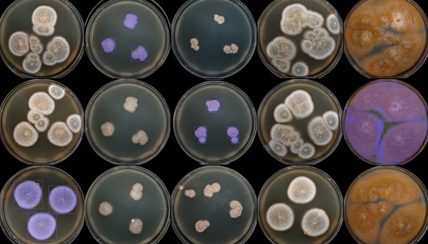
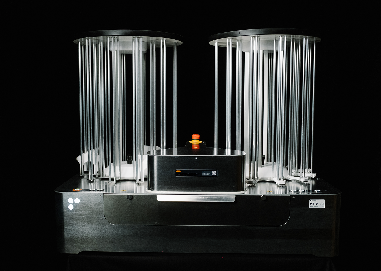
P.S. See this case study of how 21st BIO used UV mutagenesis to boost recombinant protein production in Aspergillus oryzae. With the Reshape Imaging System and AI-powered analysis, they quickly identified mutant strains with brighter fluorescence - signaling a significant jump in protein yield. The results show how pairing classical strain improvement methods with modern imaging can accelerate industrial biotechnology breakthroughs.
Sources:
Chalfie, M., Tu, Y., Euskirchen, G., Ward, W. W. & Prasher, D. C. Green fluorescent protein as a marker for gene expression. Science 263, 802–805 (1994). https://doi.org/10.1126/science.8303295
Fire, A., Xu, S., Montgomery, M. K., Kostas, S. A., Driver, S. E. & Mello, C. C. Potent and specific genetic interference by double-stranded RNA in Caenorhabditis elegans. Nature 391, 806–811 (1998). https://doi.org/10.1038/35888
Franke-Fayard, B., Trueman, H., Ramesar, J., Mendoza, J., van der Keur, M., van der Linden, R., Sinden, R. E., Waters, A. P. & Janse, C. J. A Plasmodium berghei reference line that constitutively expresses GFP at a high level throughout the complete life cycle. Mol. Biochem. Parasitol. 137, 23–33 (2004). https://doi.org/10.1016/j.molbiopara.2004.04.007
Talman, A. M., Blagborough, A. M. & Sinden, R. E. A Plasmodium falciparum strain expressing GFP throughout the parasite's life-cycle. PLOS ONE 5, e9156 (2010). https://doi.org/10.1371/journal.pone.0009156
Choe, C. P., Choi, S.-Y., Kee, Y., Kim, M. J., Kim, S.-H., Lee, Y., Park, H.-C. & Ro, H. Transgenic fluorescent zebrafish lines that have revolutionized biomedical research. Lab. Anim. Res.37, 26 (2021). https://doi.org/10.1186/s42826-021-00098-5